Anatomy of Kidneys
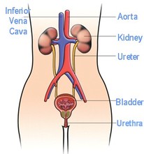
- Kidneys are a pair of red-brown organs attached to the back of the abdominal cavity, surrounded by a thick protective layer of fat and connective tissue. On a human, if you place your hands on your hips, your thumbs are in the approximate position of your kidneys.
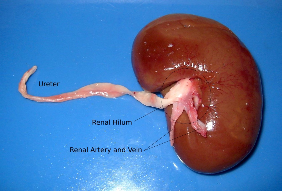
- Kidneys are supplied with blood at arterial pressure by renal arteries which branch off the abdominal aorta.
- Blood leaves the kidneys through the renal vein, into the inferior vena cava.
- The kidneys are important for blood filtration, excretion and osmoregulation.
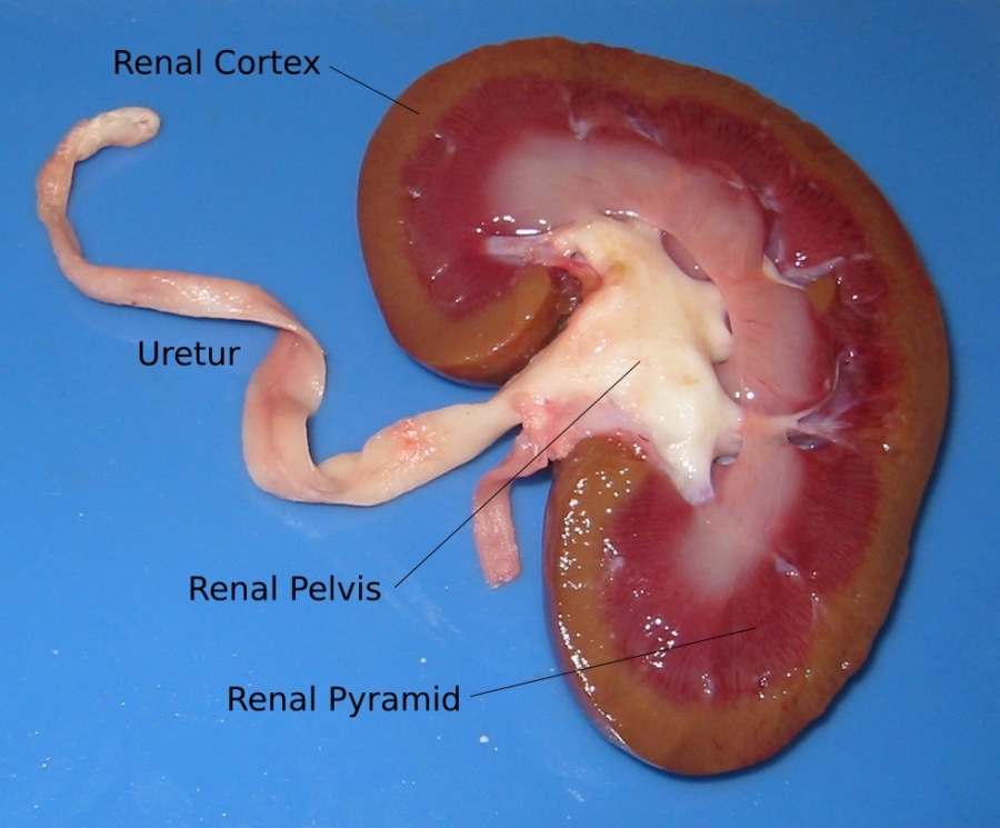
Kidney Structure
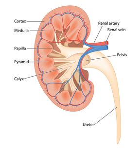
- The kidneys are made up of millions of nephrons, which act as tiny filtering units.
- The cortex is the dark outer layer. This has a high density of capillaries as it is the site of blood filtration.
- The medulla is the lighter area inside the cortex. This contain nephron tubules which make the kidney pyramids and collecting ducts.
- The pelvis is the innermost part of the kidney. It collects urine before it passes down to the bladder.
- The ureter is a tube which connects the kidney to the bladder.
- The bladder is a muscular sac for holding urine.
- The urethra is a tube which allows for urine in the bladder to be excreted from the body.
Nephrons
- Blood is filtered in the nephrons, and the majority of the filtered material returns to the blood.
- It removes nitrogenous waste, and balances mineral ions and water levels in the blood.
- Each nephron is about 3cm long, and there are 1.5 million per kidney. This provides the body with several kilometres for reabsorption of water, glucose, salts etc.
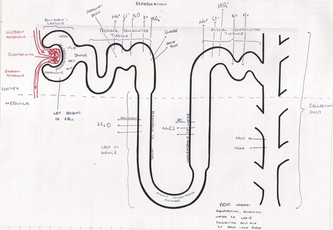
- Bowerman's capsule: a cup shaped structure containing the glomerulus where ultrafiltration takes place.
- Glomerulus: a tangle of capillaries in which the pressure forces all solutes in the blood plasma to be forced through the capillary walls. This includes ions, amino acids, glucose, urea, water. Proteins and erythrocytes do not pass through as they are too large.
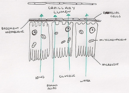
- Proximal Convoluted Tubule: First coiled region of the tubule, where products needed in the blood (ions, glucose, amino acids etc) are reabsorbed into the blood.
- Loop of Henle: A long loop of tubule which spans the cortex and medulla, used to concentrate the urine. A salty environment is created in the medulla in order to cause water to osmose of water out of the nephron on the falling limb, and the impermeable rising limb allows salts to diffuse out maintaining salty conditions.
- Distal Convoluted Tubule: Second coiled region of the tubule, where osmosis and diffusion of solutes occurs in order to fine tune the water potential and pH of the blood. Antidiuretic Hormone (ADH) affects the permeability of the distal convoluted tubule.
- Collecting Duct: Urine travels through the collecting duct down to the pelvis. More fine tuning occurs, as ADH creates aquaporins to allow the exit of excess water.
Ultrafiltration in the Glomerulus and Bowman's Capsule
- The glomerulus is supplied with blood from a comparatively wide afferent renal artery, but leaves through a narrower efferent renal artery. This means the blood in the glomerulus is under very high pressure.
- This causes the contents of the blood to be forced through the capillary wall, like a sieve, then through the basement membrane (a second 'sieve' made of collagen fibres and protein) into the nephron.
- These sieve like structures are important as they do not allow cells, large proteins or platelets to pass into the nephron.
- The cells in the wall of the bowman's capsule contain cells called podocytes, which have extensions called pedicels wrapped around the capillaries meaning any cells, large proteins or platelets which leave the capillary walls don't enter the tubule.
- Filtrates include water, amino acids, glucose, ions and importantly: urea and other nitrogenous waste products.
|
Reabsorption in the Proximal Convoluted Tubule
|
The Loop of Henle
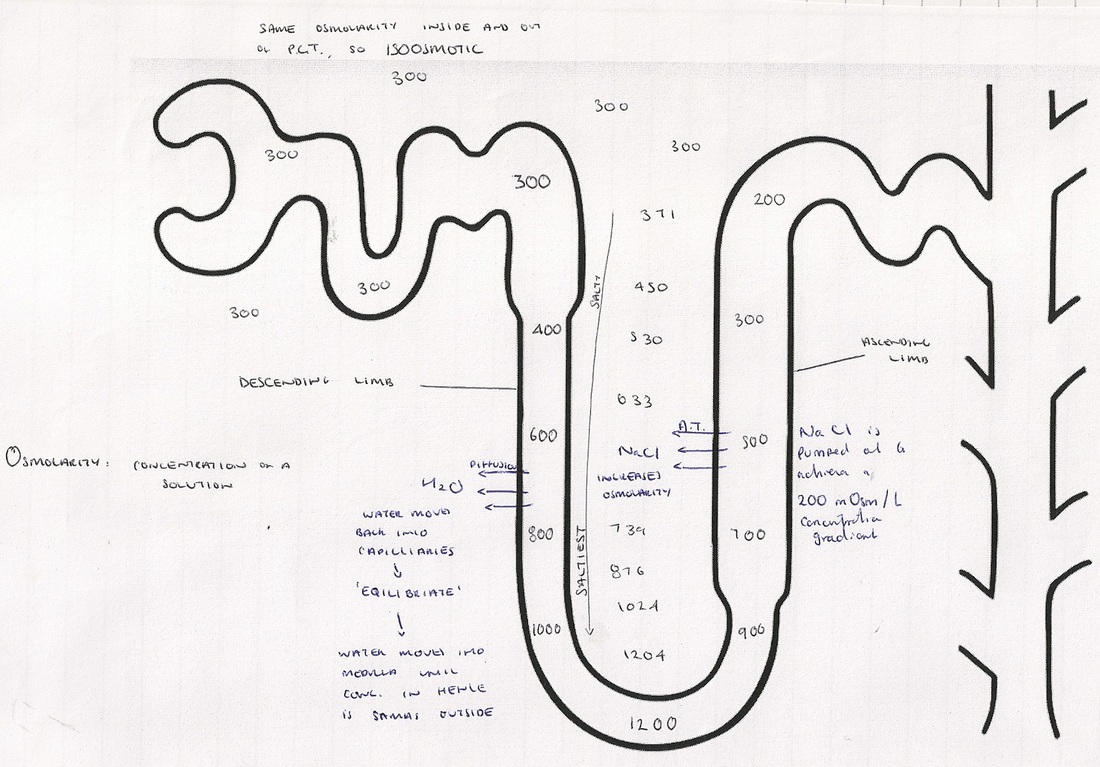
- The loop of henle allows mammals to produce urine which is more concentrated my their own blood, meaning water can be conserved while removing waste.
- Different sections of the loop of henle have different permeabilities.
- Active transport of sodium and chloride ions is used to create a concentration gradient so water moves from inside the nephron back into the capillary network. The whole process depends on high concentration of solute in the medulla creating a low water potential.
Osmolarity: Concentration of a solution expressed as total number of solute particles per litre.
Isosmotic: To have the same osmolarity as another fluid.
Equilibrate: To bring into a state of equilibrium.
Interstitial Fluid: Fluid which surrounds tissue cells.
Countercurrent Multiplication in the Loop of Henle
- Before moving into the loop of henle the filtrate in the nephron is isosmotic with the blood.
- Sodium and chloride ions are pumped out of the ascending limb to create a water potential gradient.
- The filtrate moves down the falling limb, and water osmoses out due to the water potential gradient. The ideal gradient is 200 mOsm.
- Water in the falling limb equilibrates with the interstitial fluid, by passively diffusing into the medulla. This does not change the osmolarity of the interstitial fluid.
- When the filtrate moves around the loop of henle, the ideal 200 mOsm gradient is lost, so steps 3 and 4 are repeated.
- After enough filtrate has passed through, the interstitial fluid at the bottom of the loop has reached a maximum osmolarity of ~1200 mOsm.
Osmoregulation and the Distal Convoluted Tubule
- Osmoregulation is the control of water and salt levels in a fluid, which in this case is blood.
- High levels of ADH are released when you are dehydrated, and cause the distal convoluted tubule walls to become more permeable to water, meaning more returns to the blood and the urine becomes more concentrated. This way we conserve water.
- There are many mitochondria in the tubule cell walls, allowing salts to be actively transported out of the nephron into the blood when required. Sodium ions are actively pumped out, and chloride ions follow passively down an electrochemical gradient.
- The urine enters the collecting duct which transports it down through the medulla to the pelvis.
- The urine becomes more concentrated as it passes through the medulla, as the salty conditions created by the loop of henle mean water osmoses out down a potential gradient.
- ADH also controls the permeability of the collecting duct, and creates aquaporins in the membrane which allow additional water to escape through.