Membrane Structure:
- All membranes in a cell have the same basic structure, which compartmentalises organelles from its external environment.
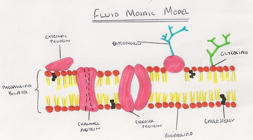
- Formed from a phospholipid bilayer, with the hydrophobic tails pointing towards each other, and the hydrophilic heads pointing outwards.
- Most accurate current model is the fluid mosaic model, which suggests the phospholipids are free to move within their layer.
Cell Membrane Components:
- Intrinsic Proteins: Proteins embedded in the phospholipid layers, held in place by hydrophobic R groups at the surface of the protein, which interact with the hydrophobic tails of the phospholipids. Can be in one or both sides.
- Channel Proteins: provide a hydrophilic tunnel that allows diffusion of polar molecules and ions down a concentration gradient.
- Carrier Proteins: used in diffusion, but mainly for active transport where ATP changes the shape of the protein allowing molecules to pass through.
- Glycoproteins: Proteins with a saccharide attached, embedded in one layer of the membrane and are receptors of chemical signals, such as for neurotransmitters in the synapses or for insulin to affect uptake of glucose in the cell.
- Extrinsic Proteins: Proteins which are loosely bound to the exterior of the membrane, by weak hydrophilic interactions with the phosphate heads.
- Glycolipids: Lipids with a saccharide attached, and are cell markers which can be recognised by cells in the immune system as self or non-self.
- Cholesterol: A sterol which regulates the fluidity of membranes. Cholesterol molecules are positioned between phospholipids, with the hydrophobic/hydrophilic ends interacting and pulling them together, adding stability to membranes.
- Many proteins involved in reactions vital for life, such as ATP synthase enzyme on the interior lipid layer, are found in the bilayer.
