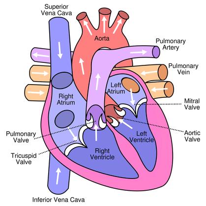
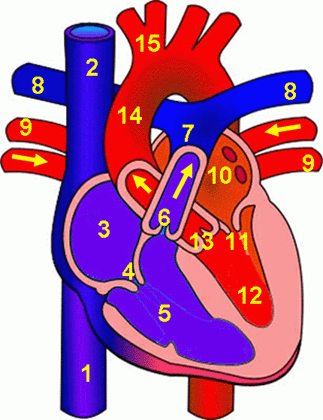
The Human Heart:
- Blood pumping organ, which pumps blood to the lungs and around the rest of the body.
- Every part of the heart has its own purpose:
- Aorta: major artery which takes blood to the body.
- Pulmonary Artery: major artery which takes blood to the lungs to be oxygenated.
- Pulmonary Veins: major veins which bring oxygenated blood from the lungs back to the heart.
- Vena Cava: Major vein which brings deoxygenated blood from the body back to the heart.
- Right Atrium: small chamber of the heart which forces deoxygenated blood into the right ventricle.
- Right Ventricle: large chamber of the heart which forces deoxygenated blood to the lungs.
- Left Atrium: small chamber of the heart which forces oxygenated blood into the left ventricle.
- Left Ventricle: large chamber of the heart which forces oxygenated blood around the body.
- Tricupsid and Bicuspid (mitral) Valves: valves which stop backflow of blood into the atria.
- Semilunar (Pulmonary/Aortic) Valves: valves which stop backflow of blood into the ventricles.
- Tendinous Cords: stop valves from being turned inside out.
- Septum: wall between the two chambers of the heart which stops oxygenated and deoxygenated blood mixing.
- Cardiac Muscle: the muscle that the heart is made of, which beats rhythmically and does not fatigue like other muscle. It is myogenic (self stimulating).
- Coronary Artery: Artery which supplies the cardiac muscle with blood.
|
| ||||
Movement of blood through the heart:
Right Side
Right Side
- Deoxygenated blood enters right atrium through vena cava at low pressure.
- Pressure builds in atrium until tricuspid valve is forced open, and blood flows into right ventricle.
- When both the atrium and ventricle are full, the atrium contracts forcing extra blood into the ventricle, stretching the ventricular walls.
- The tricuspid valve closes, the ventricle contracts and the pulmonary semilunar valve is forced open and blood passes through out to the lungs.
- Pulmonary semilunar valve closes.
- Oxygenated blood from the body enters the left atrium through the pulmonary vein.
- Pressure builds in atrium into bicuspid valve is forced open, and blood flows into left ventricle.
- When both the atrium and ventricle are full, the atrium contracts and forces extra blood into the ventricle, stretching the ventricular walls.
- The bicuspid valve closes and the ventricle contracts, forcing the aortic semilunar valve open and the blood passes out to the lungs.
- The aortic semilunar valve closes.
- Both sides of the heart fill and empty simultaneously.
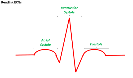
Key Terms:
Diastole: Where the cardiac muscle is in a relaxed state.
Systole: Where the cardiac muscle is contracting.
- In diastole, the chambers fill, the volume and pressure in the heart increases, but the pressure in the arteries is at its minimum.
- In systole, blood is forced out of the chambers. Volume decreases rapidly and pressure increases rapidly as chambers contract. Pressure in arteries is at a maximum, due to pulse surge.
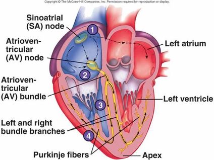
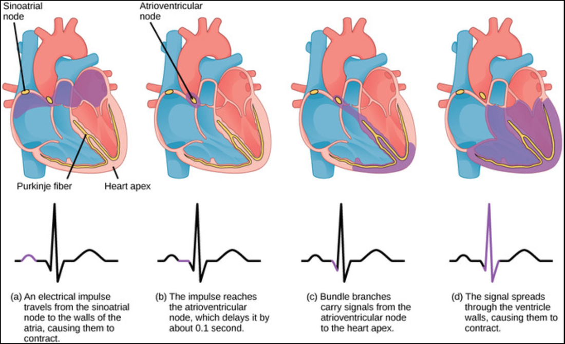
Electrical System of the Heart:
Graphical Representations of Changes in the Heart:
- The myogenic cardiac muscle stimulates its own wave of excitation which travels through the purkyne fibres and causing the chambers of the heart to contract.
- The different part of the electrical system of the heart have different purposes:
- Sino-Atrial Node (SAN): triggers the wave of excitation.
- Atrio-Ventricular Node (AVN): delays the impulse, transmits it to the bundle of his
- Bundle of His: Lots of purkyne fibres that penetrate the septum.
- Purkyne Fibres: conductive tissue which carries the impulse.
- Apex: the bottom of the heart.
- Wave of excitation begins in the Sino-Atrial node (pacemaker). This causes the atria to contract and initiates the heartbeat. Insulating tissue stops the wave of excitation being spread immediately to the ventricles.
- The wave of excitation triggers the Atrio-Ventricular node (which causes a delay) to transmit the impulse to the bundle of his.
- The impulse travels though the septum in the bundle of his, until it splits into two branches of purkyne fibres at the apex.
- The purkyne fibres transmit the wave of excitation to the ventricles causing them to contract, starting at the apex.
Graphical Representations of Changes in the Heart:
| Download the PowerPoint for a step-by-step breakdown of the above! |
Electrocardiograms:
- Measure the spread of the wave of excitation through the heart.
- Electrodes attached to the skin pick up tiny electrical differences which result from the changes of the heart.
- Can be used to detect heart abnormalities such as:
- Tachycardia: An abnormally fast heartbeat.
- Bradycardia: An abnormally slow heartbeat.
- Ectopic Heartbeat: Extra heartbeats out of the normal rhythm.
- Atrial Fibrillation: Where the atria contact rapidly and inefficiently.
- Arrhythmia: general name to any abnormal rhythm of the heart.
